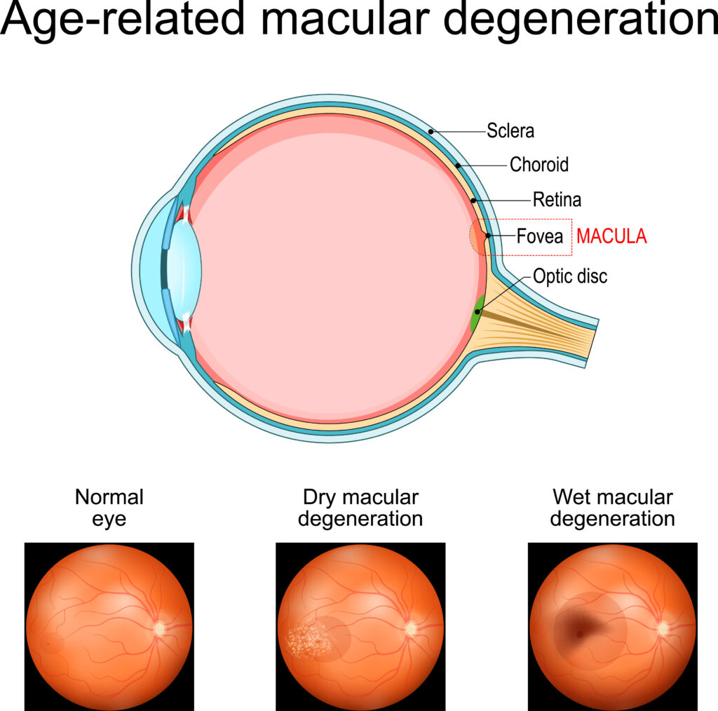
Eye doctors take pictures of the back of the eye, known as fundus photography, to get a clear view of the retina. The retina is absolutely integral to eye health and our life long vision because it contains structures that allow our brain to see. The retina is unique to the body because it is an extension of the brain, containing the optic nerve (the second of the twelve cranial nerves), many blood vessels (arteries and veins) as well as photoreceptor cells (rods and cones) that convert light into electrical signals.
Some of the most important things an Optometrist looks for when they review imaging of the retina is macular degeneration, glaucoma, blood vessel blockages, bleeding, swelling, retinal holes, tears or even retinal detachments. While there are many things that can go wrong with retinal health, it is also possible for our eye doctor to see signs of systemic disease by viewing your retinas. Since the retina is the only place in the body that we can directly see nerves and blood vessels, having your retinas examined can signal signs of diseases like diabetes, multiple sclerosis, high blood pressure, high cholesterol, autoimmune conditions and many more.
As imaging of the retina has progressed over time, the technology has improved significantly from being a simple camera flash. At Inspired Eyes Optometry, we are passionate about providing state of the art technology and the highest standard of care for our patients. Because of this, we use the Optos Monaco to provide us with the best quality images to assess the retina accurately, efficiently and (in many cases) conveniently, without the use of dilating drops at all!
The Optos Monaco is an advanced imaging device that combines multiple imaging technologies into one device. The retinal scans included in our Vision Wellness Assessments include:
- Ultra Wide-Field Retinal Imaging: High-resolution, wide-angle images of the retina, allowing for a comprehensive view in a single shot. Where an older generation camera can view 30-60 degree view, the Optos captures several times that area at 200 degrees, even without dilating drops. We like to refer to this as like “google maps” for your retina.
- Optical Coherence Tomography (OCT): This feature provides cross-sectional images of the retina, enabling detailed examination of the cellular layers. Even the earliest signs of conditions like macular degeneration can be detected with this scan that we perform at every Vision Wellness Assessment eye examination. We like to refer to this as like “google street view” for your retina.
- Fundus Autofluorescence Imaging: Non-invasive imaging technique that captures the natural fluorescence of the retina. This method provides important information specific to the health of the retinal pigment epithelium (RPE) and photoreceptors cells.
- Optic Nerve Head Scan: A crucial test that monitors the appearance of the optic nerve for patients at risk of glaucoma.
At Inspired Eyes Optometry, Vision Wellness is extremely important to us. We offer modern, convenient imaging as part of all of our eye examinations, as well as a review of those images so that patients can feel confident in their eye health. Accurate images are crucial because they allow us to detect even small changes to the health of your eyes over time.
This helps in diagnosing and monitoring various eye conditions, such as diabetic retinopathy, macular degeneration, and glaucoma. The images provide a detailed record over time, allowing the doctor to track any changes in your eye health. It’s a key part of a comprehensive eye exam!
CALL 250-862-5900Book My Appointment
Dr. Daniel Walker, OD, FOWNS, FCOVD
Dr. Walker is accepting new patients and welcomes his returning patients alike. Click on the “Book Appointment Now” button below to book a Vision Wellness Assessment appointment. We look forward to seeing you!


0 Comments